About Us
Nuclear Medicine is a branch of medicine that uses radioactive substances to diagnose various diseases and treat malignant & benign conditions. Nuclear medicine applications span a broad spectrum, ranging from risk assessment to diagnosis and treatment monitoring to radionuclide therapies. Modern nuclear medicine plays an essential role in achieving Personalized or Precision medicine, allowing both diagnosis and the selection of treatment tailored to the individual patient’s condition or predisposition towards disease.
Our Services
Initial staging of various cancers
Detection of Unknown Primary
Treatment response assessment
Restaging
Surveillance
PET CT based Radiotherapy planning,
PET CT guided biopsy
Various PET CT tracers available with us are
FDG PET-CT Whole body
F-18 NaF bone scan
F18PSMA PET- CT Whole body
F18DOPA PET- CT Whole body
Ga68 FAPI (IV) Whole body
![]() To assess myocardial viability, which aids the cardiologist and cardiothoracic vascular surgeon in deciding on management in CAD.
To assess myocardial viability, which aids the cardiologist and cardiothoracic vascular surgeon in deciding on management in CAD.
FDG PET CT is used in
Various types of Dementias
Auto-immune encephalitis
Epilepsy
Psychiatric disorders
F18 DOPA is used in disease assessment and monitoring treatment of Parkinson’s disease.
For disease assessment and monitoring treatment in
Large and small vessel vasculitis
Rheumatoid arthritis
IgG4 RD spectrum disorders
Inflammatory myopathy
Adult Still’s disease
Relapsing polychondritis
Polymyalgia rheumatica
Spondyloarthropathy
Sarcoidosis
FDG PET CT in carcinoma thyroid, medullary carcinoma of the thyroid, high-grade neuroendocrine tumors, paragangliomas
Ga68 DOTATATE in Medullary carcinoma of the thyroid, Neuroendocrine tumors, Pheochromocytomas, paragangliomas
Ga68 exendin in insulinomas
F18 DOPA in parathyroid tumors, Pheochromocytomas, paragangliomas
Osteomyelitis
Fever of unknown origin
Tuberculosis
Other Granulomatous infections

Fundamental Advantages of Nuclear Medicine
Diagnostic Nuclear medicine involves using small yet traceable amounts of radioactive substances to image and/or measure an organ’s global or regional function. The extent to which a radiopharmaceutical is absorbed, or “taken up,” by a particular organ or tissue can be quantified to determine the level of function of the organ or tissue being studied. Among the different types of radionuclides available, the type of radionuclide used will depend on the kind of study and the body part being studied.
The radioactive tracer (radiopharmaceutical) is given to the patient by intravenous injection, orally, or by other routes depending on the organ and the function to be studied. The tracer substance uptake, turnover, and/or excretion is then analyzed with a gamma camera, positron emission tomography (PET) camera, or another instrument, such as a simple stationary radiation detector. The uptake of the tracer is generally a measure of the organ function or metabolism or the organ blood flow. The acquisition of the images will be followed by image interpretation and quantification.
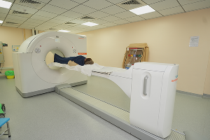
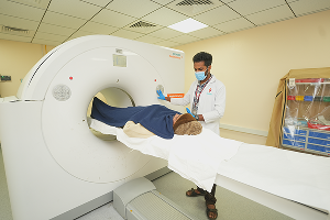
Our Facilities
Unlike other forms of imaging, a PET scan shows molecular activity and helps doctors identify diseases in the earliest stages. For this reason, a PET scan is a highly reliable tool for detecting disease processes. PET CT combines two different imaging technologies in a single scan.PET measures the body’s biological processes, and CT generates high-resolution cross-sectional images of the body’s anatomy. By combining both sets of information into a single three-dimensional hybrid image, metabolic processes are mapped accurately in all spatial dimensions.
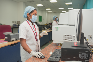
Imaging with Gamma Camera involves preparing and injecting radiopharmaceuticals that specifically trace the function of the Organ of Interest. Radiopharmaceuticals are made in the well-equipped Hot Lab in the Department of Nuclear Medicine and Molecular Imaging. These specific radiopharmaceuticals are administered to patients, and images are acquired. Static, dynamic, and parametric images can be obtained under a Gamma Camera.
![]()
Myocardial Perfusion Scan visualizes the distribution of tracer uptake in the heart muscle, which reflects regional blood flow in different coronary artery territories. This can be performed with 201T1/ 99m Tc- Sestamibi / Tetrofosmin.
Rest Injection Scan indicates whether the heart muscle is viable or scarred due to prior attacks. Stress (Exercise/Pharmacological) Scan reveals inducible ischemic perfusion defect corresponding to significant coronary disease.
Advanced Cardiac Analysis with Quantitative Perfusion
Spect and gated SPECT software provides the following Information
Universal slice review
Bullseye plot
Cardiac ejection fraction
Wall motion abnormality
Myocardial viability
Clinical Indications
Detection of Coronary Artery Disease
Chest Pain Evaluation – Stress Test with uninterpretable ECG
Assessment of borderline Coronary Stenosis seen in Angio
Assessment of Myocardial Viability
Cardiac fitness for non-cardiac surgery
A bone scan is a diagnostic imaging study that records the distribution of a radioactive tracer in the skeletal system. It is the most sensitive study available to pick up any pathology of the skeleton.
Clinical Indications
Metastatic Bone lesions
Primary Bone lesions
Osteoid Osteoma etc.
Infection/Inflammation
Osteomyelitis
Sacroilitis
Avascular Necrosis
Trauma Stress fracture
Metabolic Bone Disease
Bone pain evaluation – low back ache evaluation
Renal scans are performed to evaluate the kidney’s differential renal function, extraction, and excretion. Information about Glomerular Filtration Rate (GFR), tubular function, and cortical morphology can be obtained by such scans.
Clinical Indications
DTPA Renal scan
Split renal functions
GFR estimation
Evaluation of hydronephrosis
PUJ Stenosis
Renovascular Hypertension
Transplant evaluation
DMSA Cortical scan
Pyelonephritis
Urinary tract infection
Reflux disease
Ectopic Kidney
Voiding Cystography
Detection & follow-up of ureteric reflux
![]()
Brain Perfusion SPECT for diagnosing:
![]()
Assessment of stroke
![]()
To detect Epileptic focus
![]()
To evaluate dementia
![]()
OTHER INDICATIONS
![]()
Thyroid Scan to evaluate palpable thyroid nodule, midline neck swelling, and thyromegaly & toxic goiters
![]()
Radio Iodine Scan for post-operative thyroid cancer
![]()
Lung Ventilation Perfusion Scan for pulmonary embolism
![]()
Radionuclide Ventriculography for LV function & Ejection fraction
![]()
Isotope Venography for DVT
![]()
Scinti-mammography for doubtful breast lesion.
Hepatobiliatobiliary scan
To diagnose acute cholecystitis
To assess Gallbladder dysfunction
To study bile drainage, atresia, and post-OP cases
Liver & Spleen scan
To evaluate cirrhosis
Buddchiari, Nodular Hyperplasia, and tumors
Others scan
Hemangioma of the Liver
GI bleeding
Ectopic Gastric Mucosa(MECKEL’S DIVERTICULUM)
Esophageal transit in dysphagia
GE Reflux scintigraphy
Gastric Emptying in Gastroparesis, post-OP states, dysmotility, etc.
Nuclear medicine therapy or radionuclide therapy involves the administration into the body of radiopharmaceuticals, safe amounts of radioactive material used for both treatment purposes. Organ or tissue-specific molecules can be used safely to carry radioactive materials inside the human body to acquire more accurate images of tumors and to target and eliminate cancer cells more effectively. This method of combining therapeutic and diagnostic uses of radiopharmaceuticals is called theranostics.
Radioiodine (I-131) therapy
I-131 MIBG therapy for Pheochromocytomas and paragangliomas
Metastatic bone pain palliation
In prostate cancer
In Neuroendocrine tumors -PRRT
In Hepatocellular carcinomas and liver metastasis
Radiosynovectomy
![]() Radioguided surgeries utilize a gamma probe supplemented with SPECT imaging after administering radiopharmaceuticals in breast cancer, parathyroid adenomas, melanomas, etc.
Radioguided surgeries utilize a gamma probe supplemented with SPECT imaging after administering radiopharmaceuticals in breast cancer, parathyroid adenomas, melanomas, etc.

Dr. Anitha is the lead specialist in the department of Nuclear Medicine at Vydehi Institute of medical sciences and research center. With the experience of more than a decade, Dr. Anitha has been providing prime-grade nuclear medicine-related diagnosis and treatment services.
Education
Dr. Anitha is an MBBS graduate from Government Kilpauk Medical College, Chennai.
Completed her DNB in Nuclear Medicine from Seth G.S Medical College and K.E.M Hospital, Mumbai.
Highlights
Dr. Anitha is a certified RSO (II). Reported more than 5000 PET-CT cases to date. Core interest is Theranostics. Certification for ‘the Thyroid preceptorship at AIIMS under Dr.Chandrasekhar Bal’ in 2016.
Accomplishments
Dr. Anitha published an article entitled “Tc-99m EthylenediCysteine and Tc-99m DiMercaptoSuccinic Acid scintigraphy – comparison of the two for the detection of scarring and Differential Cortical Function in urinary tract infections” in the Indian Journal of Nuclear Medicine and at the international conference conducted by EANM 2016, Barcelona, Spain are one of her remarkable works. She also presented the “ROLE OF THREE PHASE BONE SCINTIGRAPHY AND SPECT/CT IN VARIED PRESENTATION OF OSTEOID OSTEOMA” poster at the 47th Annual conference of the Society of Nuclear Medicine, India.