


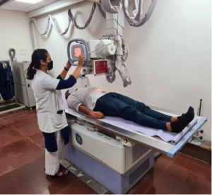
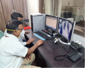
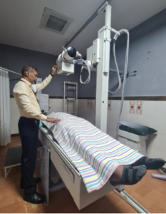
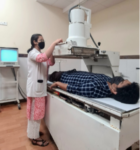
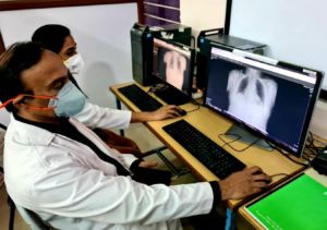
Radiography and Fluoroscopy use X-ray technology to diagnose disease and injury inside the body. Radiography is used to examine the chest, abdomen, skull and long bones. A specialized X-ray machine with fluoroscopy is used to conduct barium examinations of GI tract & iodine examinations of the genito-urinary system which include cystourethrogram (MCU, RGU), Intravenous Pyelogram (IVP) and Hysterosalpingogram (HSG). All these radiographic and fluoroscopy procedures are routinely done in our hospital.
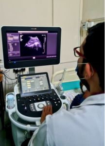
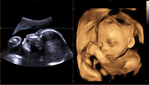
Ultrasonography uses high-frequency sound waves to create an image. This technique does not involve ionizing radiation and is very safe for pregnant women and young children. Since images are obtained in real time, our radiologists can obtain detailed examination of both structure and function. Common examinations include evaluation of abdominal organs, neck, breast, musculoskeletal structures, male and female pelvis, antenatal scans, new-born brain, blood vessels, lungs and heart. Advanced 3D ultrasound is used in fetal imaging, where in depth information of the baby’s well-being helps to detect anomalies with high precision. Doppler ultrasound is used to assess the motion of blood within the vessels which aids in diagnosis of a wide range of vascular pathologies.
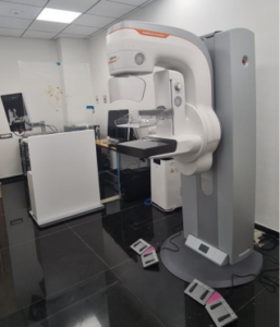
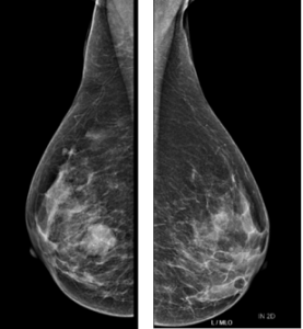
Mammography is a low-dose X-ray procedure used to produce the digital image of the breast. Mammogram can identify breast cancer long before it can be detected by a physical examination. In addition, high resolution sono-mammography is also routinely performed, which are useful for detection of very small cancers especially in younger and high-risk women. In Vydehi Hospital, we have state of art most advanced equipment with Tomosynthesis and Stereotactic Biopsy. We encourage all women to begin regular mammography screenings starting at age 40 whilst high risk group may be advised to begin at a younger age.
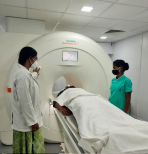
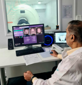
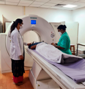
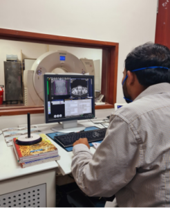
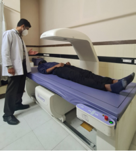
Computerized Tomography (CT) provides clear three-dimensional images of body organs and their structure. Due to the high resolution, images are clearer than conventional X-rays. The CT scans can help doctors to make an early and accurate diagnosis. While undergoing a CT scan you are required to lie still on the CT couch for a short period of time which will be moved in and out through a CT gantry. You may require an oral and / or intravenous contrast agent to enhance quality of the images. Intravenous contrast agents are iodine based that may make you feel warm for some time. At Vydehi Hospital, all routine CT scans are done with 128 slice scanners. Siemens Definition AS 128 slice spiral CT scanner is a state of art multi detector CT (MDCT) scanner which generates 128 slices in less than a second. As a result of the fast scanning & high tube capacity, very thin sections of high resolution images can be obtained quickly. CT coronary angiography, the technique for scanning of blood supply to the heart in an accurate and non-invasive manner is frequently performed in our hospital. Other CT angiography studies of brain, neck, abdomen, upper & lower limbs, musculoskeletal 3D CT studies, virtual bronchoscopy, virtual colonoscopy, multiphasic abdominal CT & CT perfusion studies are also done with precise image definition. With our high-speed scanners, routine CT studies are done with very high resolution in light speed making it ideal for very sick patients, polytrauma patients, elderly and infants or young children. The data can be transmitted in a CD for digital storage & archiving.
Magnetic Resonance Imaging (MRI) is a highly advanced, painless, non-invasive test that creates excellent high-resolution images of the internal structures and organs of your body. It is primarily used in medical imaging to demonstrate pathological or other physiological alterations within the organs. At Vydehi Hospital, we have Siemens MAGNETRON SEMPRA 1.5T MRI equipment which gives us tremendous diagnostic capabilities. This state of art equipment has commendable speed and near to nil audible noise which is more comfortable and convenient for claustrophobic patients. Our areas of expertise are Cardiac MRI, Neuroradiology (Head, Neck and Spine), MR spectroscopy, MR perfusion imaging, and Musculoskeletal, Pelvic and Abdominal MRI examinations.
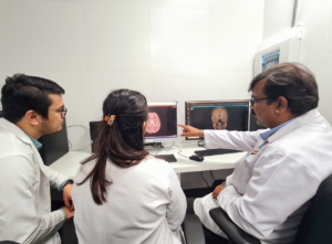
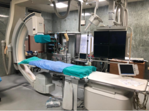
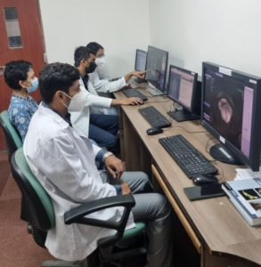
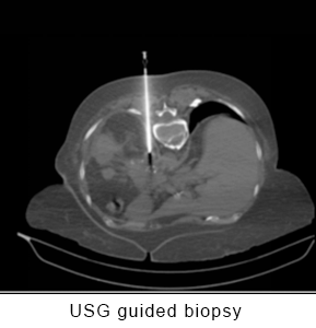
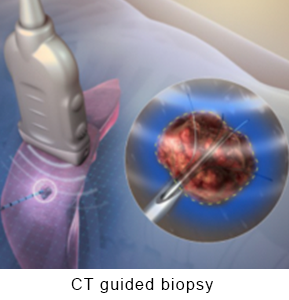
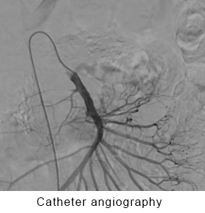
Interventional radiology is a medical specialization that involves performing a range of imaging procedures. Interventional radiologists provide distinct services in advanced procedures such as treating tumours, taking organ biopsies or placing stents by inserting tiny instruments and thin plastic tubes (catheters) into the body via an artery or vein. The images are used to guide the catheters and instruments to the exact area where the procedure or treatment is to be performed. This reduces the need for traditional (open) or keyhole (laparoscopic) surgery as treatment can be given via a small plastic tube about the size of a straw.
Sub-unit of Interventional radiology in Vydehi performs the following wide range of procedures routinely:

 Emergency Number